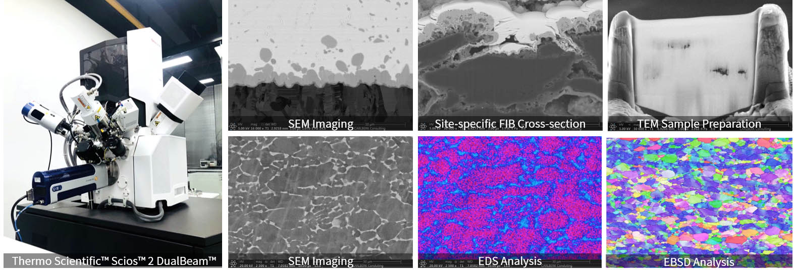
FIB/SEM
CarlBerk’s central analytical lab is equipped with a Thermo Scientific™ Scios™ 2 DualBeam™ workstation, providing outstanding sample preparation and microstructure characterization for a wide ranges of samples. The dual-beam focused-ion-beam (FIB) workstation consists of a FIB column and a scanning electron microscope (SEM) column on the same platform. In addition to traditional uses of a field emission SEM, dual beam system allowed ion imaging and site-specific cross-sectional milling to support detailed microstructure analysis. By incorporating an ultrafine tungsten manipulator probe, preparation of TEM lamella and atom probe needles could also be achieved in the FIB system.
The dual beam workstation is installed with an Ultim Max EDS detector and Symmetry EBSD detector, which allows obtaining of chemical spectra, elemental and orientational mapping with great speed and sensitivity and thus supports comprehensive analysis over materials morphology, structure, and chemistry.
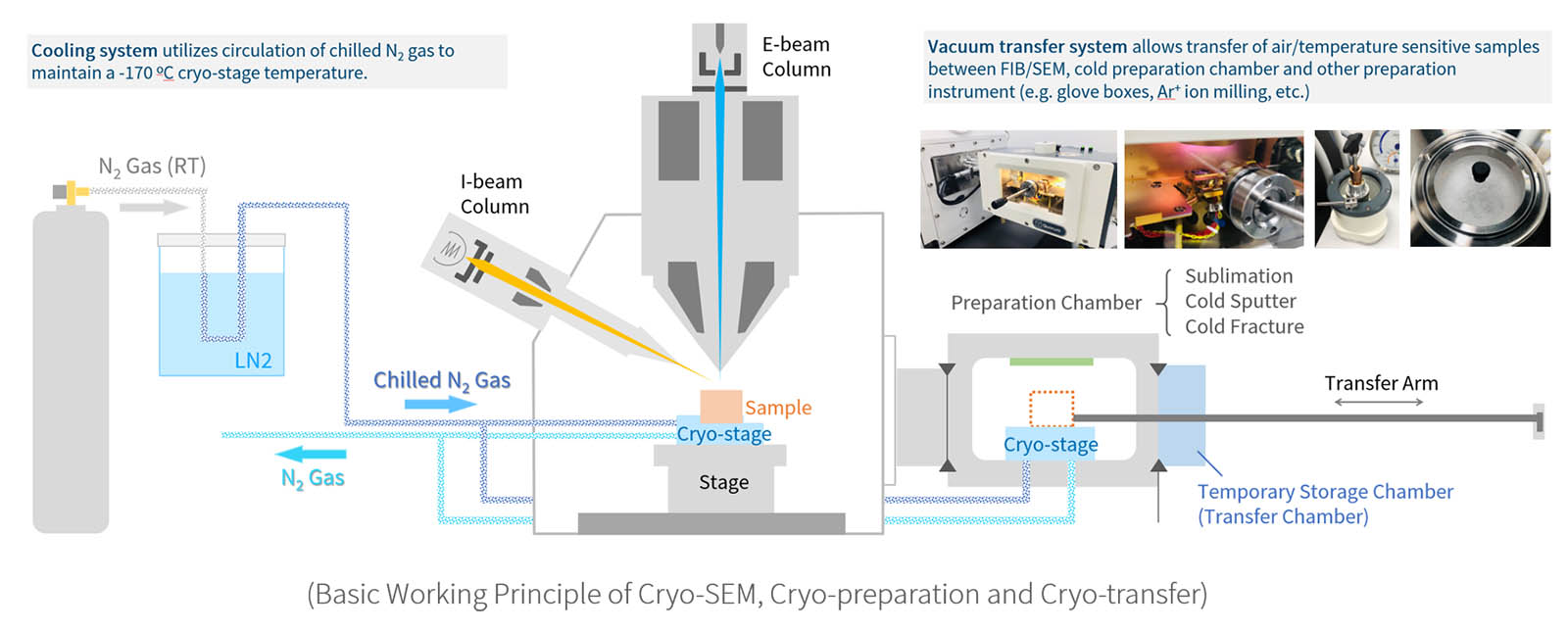
Cryo-FIB/SEM
The most fascinating part of our workstation, which makes the system really stand out from the regular FIB/SEM, is that it is equipped with a highly automated, column-mounted, gas-cooled cryo preparation and vacuum transfer system (Quorum PP3010). In this system, the sample stage is cooled to near liquid nitrogen temperature by chilled nitrogen gas and allows successful observation or processing of wet or ‘beam sensitive’ specimen. The system is also equipped with a turbomolecular pumped preparation chamber for cold fracture, sublimation, and sputter coating treatment, and a transfer system for transferring sensitive sample between SEM, preparation chamber and other instrument (e.g. glove box, cross-section polisher, etc.).
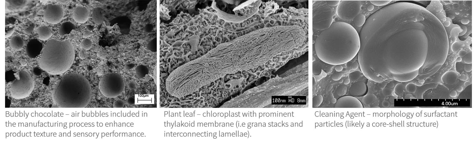
Images taken from Cryo-FIB/SEM
Cryo preparation techniques for cryo SEM and FIB/SEM are essential for the observation of biological, wet or beam sensitive specimens. Using cryo sem electron microscopy removes the need for conventional preparation methods, such as chemical fixation and critical point drying, and allows observation of specimens in their natural hydrated state.
![]()
1058 S. Shenbin Rd., Bldg. D, Suite 510, Minhang, Shanghai
(US) 1-734-709-7508 | (China) +86 137 0178 7520
© 2018-2023 CarlBerk Consulting